A Story of a Mysterious Illness and Modern Imaging Technology
Late at night, Arif suddenly began to experience chest pain. The pain was so severe that he could barely breathe. His family, alarmed, rushed him to the hospital. The doctor performed some basic tests and suspected there might be a problem with his lungs or heart. But to be sure, more in-depth examinations were needed—something that could clearly reveal the state inside his body.

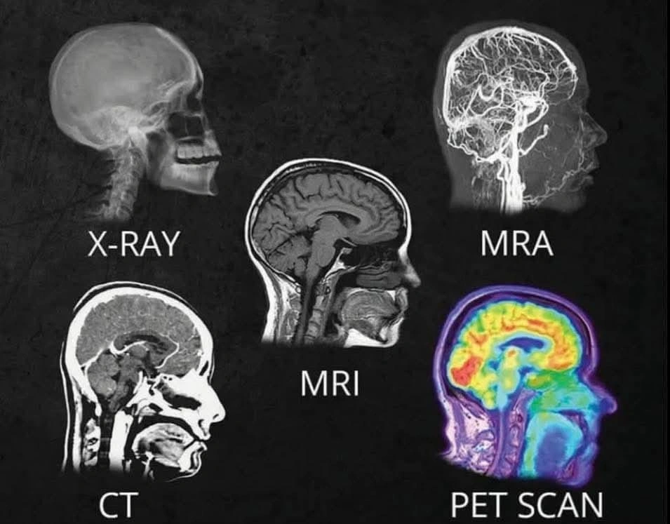
In this situation, the doctor mentioned a series of imaging technologies—X-Ray, MRA, MRI, CT, and PET scans. But how do these work? How can these technologies unlock the hidden mysteries inside our bodies? Let’s discover these vital technologies of modern medical science through a compelling story.
X-Ray: The First Eye of Medical Science
Upon arrival at the hospital, the doctor first recommended a chest X-ray for Arif. In 1895, when Wilhelm Röntgen first discovered the X-ray, it was a groundbreaking event.
X-rays are actually a form of high-powered rays that can penetrate the body and are absorbed in varying degrees depending on the tissue type. For example, bones absorb more X-rays, so they appear white in the image, whereas the lungs or soft tissue absorb less, showing up as black or grey.
📌 Key Statistics:
Approximately 3.6 billion X-ray scans are performed worldwide every year.
A standard X-ray is low-cost and takes only 5-10 minutes to complete.
After reviewing the X-ray report, the doctor said, “There are minor issues visible in your lungs, but further detailed tests are needed. Next, we need to do an MRI.”
MRI: In-Depth Observation through Magnetic Power
MRI, or Magnetic Resonance Imaging, is a technology that uses magnets and radio waves to create detailed images of the body’s soft tissues. It’s more advanced than X-rays because it doesn’t use ionizing radiation, and it’s highly effective in examining the brain, spine, joints, heart, and other soft tissues.
Inside the MRI machine, Arif felt as if he was in a spaceship! There was a humming sound all around, and the instructions from the doctor were, “Stay completely still.”
📌 Key Statistics:
Over 60 million MRI scans are performed globally every year.
An MRI scan usually takes around 30-60 minutes.
After reviewing the MRI report, the doctor said, “Everything looks fine, but to get an even clearer picture, we will do a CT scan.”
CT Scan: Creating Three-Dimensional Images of the Body
CT scan (Computed Tomography or CT Scan) is an advanced X-ray technology that creates detailed three-dimensional (3D) images of the body. Like an X-ray, it uses radiation but captures images from multiple angles, allowing doctors to analyze organs in greater depth.
To further understand the cause of Arif’s chest pain, the doctor ordered a CT angiogram, which is used to detect blockages in blood vessels. This is highly effective in assessing risks for heart disease and stroke.
📌 Key Statistics:
Over 80 million CT scans are performed annually worldwide.
A typical CT scan takes only 5-10 minutes.
After receiving the CT scan report, the doctor said, “Nothing is conclusive yet. To get more detailed information, a PET scan is necessary.”
PET Scan: Detecting Disease at the Cellular Level
PET (Positron Emission Tomography) is one of the most advanced imaging technologies, helping to observe cellular activity within the body. It is mainly used for cancer detection, diagnosing brain disorders, and analyzing heart disease risks.
For this scan, the patient is given a type of radioactive tracer, which accumulates in specific parts of the body and creates images based on cellular activity.
📌 Key Statistics:
More than 2 million PET scans are conducted globally each year.
It is used most commonly for cancer detection (90%).
Through the PET scan, doctors confirmed that Arif had no blockages in his heart; rather, the problem was muscle-related, which would heal with medication and rest.
Final Thoughts: Technology Is Saving Our Lives
When Arif returned home from the hospital, he realized that without these technologies, identifying his problem would have been very challenging. These medical imaging technologies are saving countless lives every day.
Today, 70% of diagnoses depend on imaging technologies. According to the World Health Organization (WHO), in developed countries, 120-150 out of every 1,000 people undergo at least one imaging scan per year.
So, if a doctor ever recommends any of these scans for you, there’s no need to be afraid. Instead, think about how science and technology are making your life easier and safer.
What Do You Think?
Have you ever had an X-ray, MRI, CT, or PET scan? What was your experience? Share in the comments!
affordablecarsales.co.nz


Leave a comment